Digital Dental X-Ray & Diagnostics
High-definition, low-radiation imaging that enables our specialists to see beyond the tooth surface for accurate diagnosis in Shankhamul, Kathmandu.
What is a Digital Dental X-Ray?
A digital dental X-ray is an advanced diagnostic imaging technology that utilizes ultra-sensitive electronic sensors instead of traditional chemical film. It captures crystal-clear images of your teeth, roots, and jawbone instantly on our HD clinical monitors with zero waiting time.
Why are Dental X-Rays Essential?
Over 50% of serious dental issues—such as root canal infections, bone loss from gum disease, and interdental cavities—develop beneath the surface where visual inspection cannot reach. Digital radiography enables early detection before severe pain or tooth loss occurs.
When Do You Need an X-Ray?
We rely on digital imaging across various diagnostic workflows:
-
•
Routine Check-ups Annual screening to monitor bone levels and check for unseen decay.
-
•
Unexplained Tooth Pain Pinpointing the exact root nerve source of pain or swelling.
-
•
Root Canals & Implants Mapping root canal anatomy and measuring jawbone density for dental implants.
What Digital X-Rays Reveal
•Interdental Cavities
Decay forming between tight teeth where mirrors cannot reach.
•Periapical Abscesses
Infections and cysts developing at the root tips inside the jaw.
•Bone Density Loss
Measuring bone degradation caused by untreated periodontal disease.
•Impacted Wisdom Teeth
Determining exact angles and nerve proximity of trapped teeth.
Types of Digital X-Rays
• Periapical (IOPA) X-Ray
Captures the entire tooth from crown to root apex. Essential for diagnosing root canal infections and assessing bone health.
• Bitewing X-Ray
Displays upper and lower back teeth simultaneously. The clinical gold standard for detecting decay between molar teeth.
• Panoramic X-Ray (OPG)
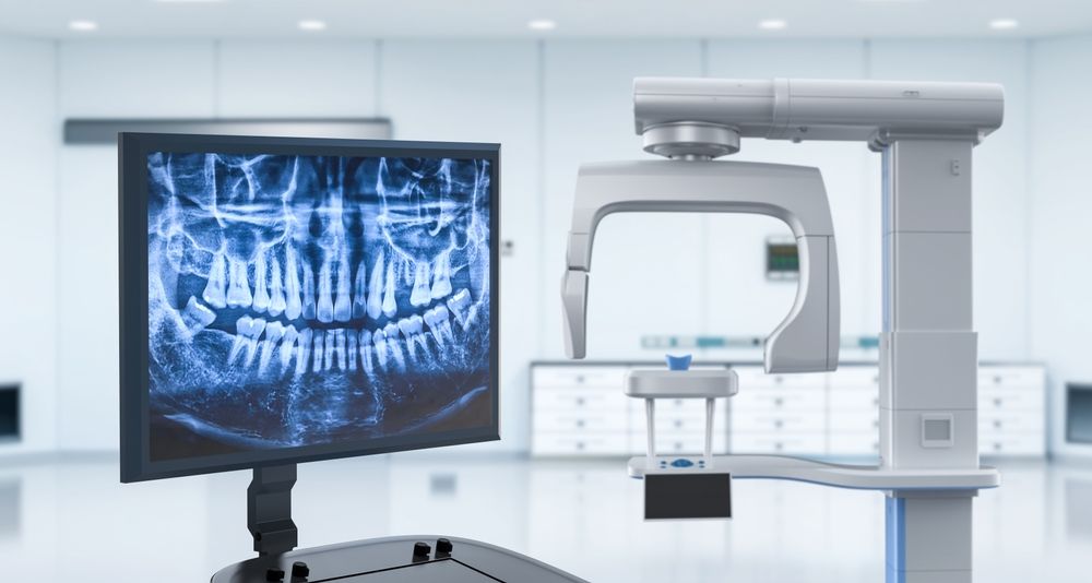
A single wide scan of the entire upper and lower jaw, joints (TMJ), and sinuses. Used for implants, wisdom teeth, and orthodontics.
Why Digital X-Rays Are Superior
Ultra-Low Radiation
Our digital sensors reduce radiation exposure by up to 90% compared to legacy film X-rays.
Instant HD Preview
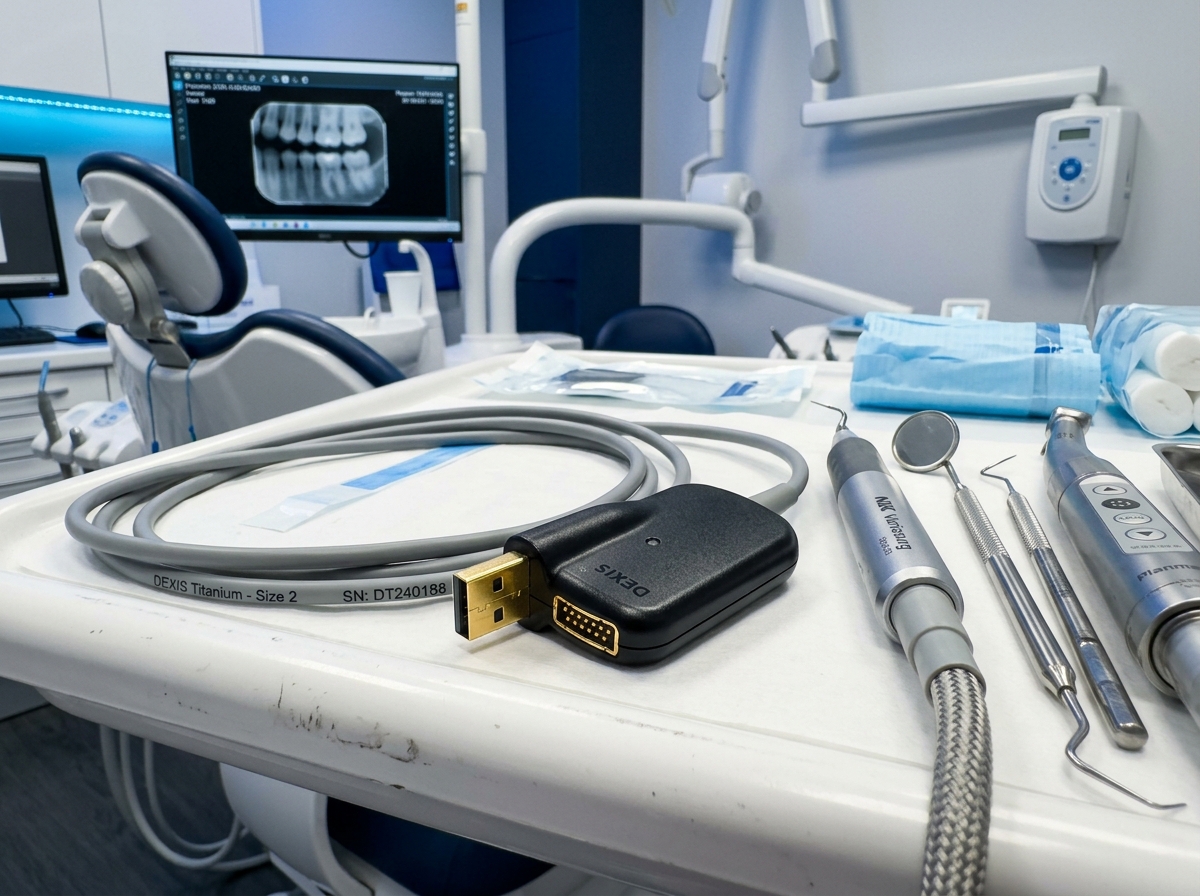
No waiting for darkroom chemicals. High-resolution images appear immediately on chairside monitors.
Enhanced Zoom & Contrast
We can magnify images and optimize contrast to reveal microscopic cracks and early-stage cavities.
Protective Shielding
We provide lead aprons and thyroid collars for maximum patient protection during every exposure.
Our Digital X-Ray Process
Clinical Assessment
Evaluating symptoms to determine needed views.
Protective Setup
Fitting lead apron and sensor positioning.
Fast Digital Capture
Painless capture in less than 3 seconds.
Chairside Review
Instant high-definition monitor display.
Transparent Review
Our dentist explains findings with you.
Targeted Care Plan
Creating a clear, personalized treatment plan.
X-Ray Diagnostics Pricing
Transparent & Fair Diagnostic Rates
Intraoral Digital X-Ray (IOPA)
Single tooth periapical root scan
Panoramic X-Ray (OPG)
Full-mouth panoramic jaw scan
* Note: The exact imaging needed is determined clinically so you only receive necessary diagnostic views.
Book Diagnostic ScanDiagnostic Suite & Technology Gallery
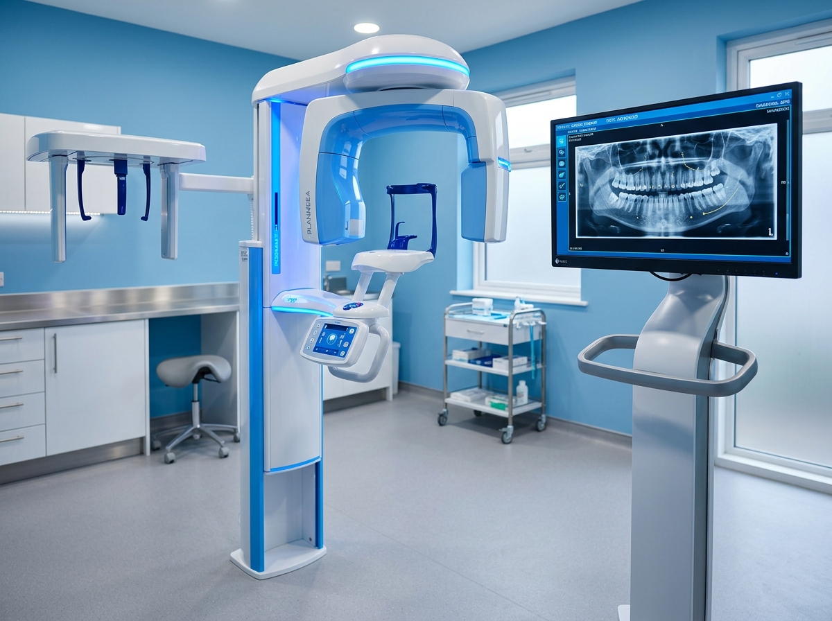
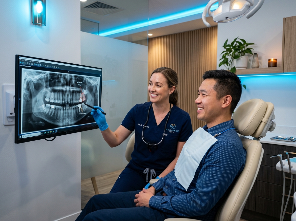
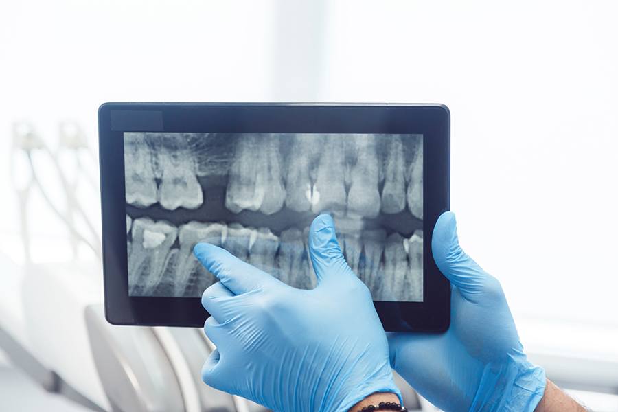
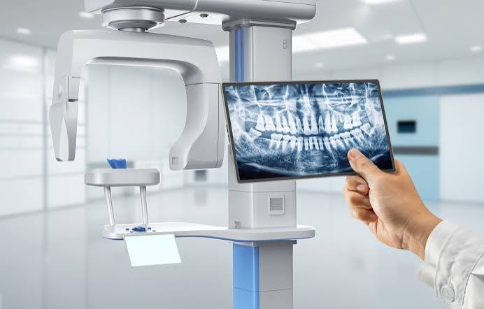
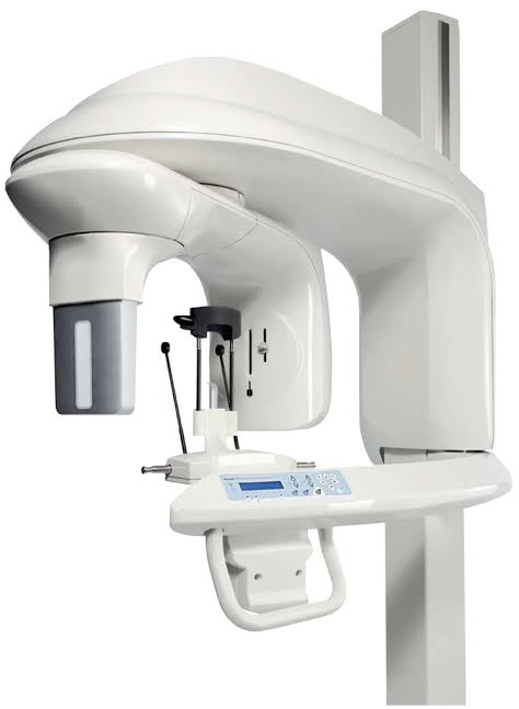
Explore the modern low-radiation diagnostic equipment and chairside digital imaging we use to safeguard your smile in Kathmandu.
Radiation Safety: The CareFirst Standard
“Accurate dentistry begins with seeing what is beneath the surface. Digital imaging gives us the clarity to save natural teeth through early, conservative interventions.”
Dr. Subash Banjade
Dental Surgeon & Clinical DirectorFrequently Asked Questions
Find answers to commonly asked questions regarding our treatments and procedures.
Related Dental Treatments
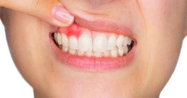
Periodontal Treatment
- Gum disease treatment
- Deep cleaning
- Healthy gums
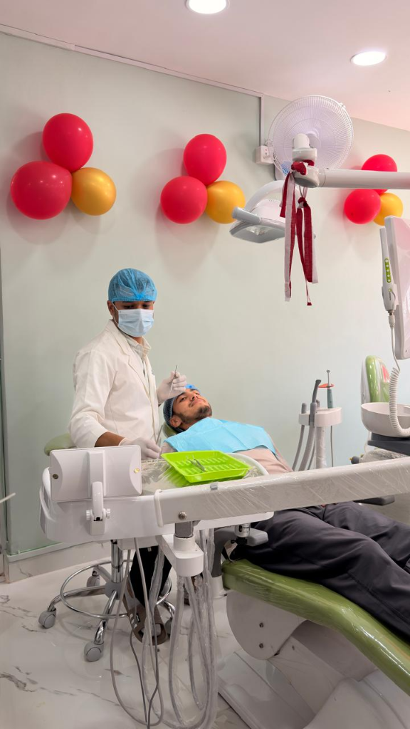
General Dentistry
- Routine check-ups
- Preventive care
- Oral health assessment
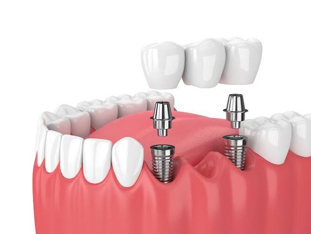
Dental Implants
- Permanent tooth replacement
- Strong & natural-looking
- Long-lasting results
Accurate Diagnosis Leads to Better Care
Experiencing tooth pain or need a comprehensive check-up? Book an appointment today for a precise digital evaluation.